A people who don’t make provision for their own sick and suffering are not worthy of civilization.”
–Daniel Hale Williams
Internal Surgery: Open-Heart Medical Milestone (1893)
On July 10, 1893, Dr. Daniel Hale Williams performed one of the world’s first successful open-heart surgeries at Provident Hospital in Chicago, Illinois. Operating without the benefits of modern antibiotics, blood transfusions, or advanced imaging, Williams successfully repaired a torn pericardium (the sac surrounding the heart), saving the patient’s life and shattering prevailing medical taboos against internal cardiac intervention.
This historic procedure solved a critical medical challenge of the late 19th century: how to treat severe penetrating chest wounds without allowing the patient to bleed to death or succumb to rampant thoracic infection.
The Innovation: The Exploratory Pericardial Repair
At the time, opening the chest cavity to operate on the heart was widely considered a death sentence by the medical establishment. Dr. Williams rejected this fatalism when James Cornish was admitted with a deep stab wound to the chest from a tavern brawl.
While the wound initially appeared minor, Cornish’s condition rapidly deteriorated as internal bleeding began compressing his vital organs. Williams made the bold decision to operate, pioneering an exploratory technique under strict antiseptic protocols.
Why the Procedure Succeeded?
- Total Antiseptic Discipline: Williams adopted the latest germ-theory practices, washing the wound cavity with a carbolic acid solution to prevent lethal post-operative infections like peritonitis or sepsis.
- Precision Suture Selection: He utilized fine catgut sutures to close the internal pericardial wound, a material that the human body could naturally absorb over time.
- Exploratory Restraint: Recognizing that the heart muscle itself had only suffered a minor superficial scratch, he left the myocardium alone and focused entirely on sealing the bleeding pericardial sac.
Key Surgical Components
The landmark operation relied on basic tools elevated by advanced technique and anatomical knowledge:
| Component / Step | Surgical Function |
| Exploratory Incision | A small opening made through the left chest wall by removing a portion of the fifth rib to grant direct access to the cardiac cavity. |
| Pericardial Irrigation | Continuous cleansing of the chest cavity with sterile salt solutions to clear pooled blood and visualize the wound. |
| Catgut Suture Application | The deliberate sewing of the torn pericardial edges while the heart actively beat beneath the surgeon’s fingers. |
| Surgical Drainage | Leaving the external chest wound partially drained to allow remaining fluids to escape, preventing fluid buildup around the lungs. |
Clinical Performance: Surviving the Unsurvivable
Medical data from the era shows that penetrating wounds to the heart carried an close to 100% mortality rate if left untreated. Williams’s swift intervention dramatically altered that statistical reality.
Patient Recovery Benchmarks:
- Untreated Cardiac Stab Wounds (1890s): Death typically occurred within minutes to a few hours from internal bleeding or cardiac tamponade.
- Cornish Post-Op (Day 7): Patient was sitting up in bed, free of fever, with regular cardiac rhythms.
- Long-Term Survival: James Cornish was discharged fully recovered 51 days after the surgery and lived for over another 20 years.
The Surgical Process
Dr. Williams executed the historic procedure in a sequence of precise, high-pressure steps:
1.Administer Anesthesia:Under 10 min.
Secure the patient and safely induce surgical anesthesia using liquid ether administered via an inhalation mask.
2.Execute Thoracic Window:Careful rib resection.
Extend the existing stab wound to the left, carefully resecting a piece of the fifth rib to create a clear physical window into the chest cavity.
3.Ligate the Internal Mammary Artery:Immediate hemorrhage control.
Locate and tie off the punctured internal mammary artery to halt the primary source of arterial bleeding into the chest.
4.Inspect and Suture the Pericardium:Active cardiac intervention.
Inspect the moving heart. Clear out internal blood clots, identify the 1.25-inch tear in the pericardium, and loop fine catgut sutures to close the membrane.
5.Close and Drain:Post-operative stabilization.
Close the external thoracic wall wounds using silk sutures while maintaining a sterile compress dressing over the surgical site.
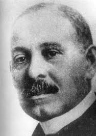
About the Surgeon: Dr. Daniel Hale Williams
Daniel Hale Williams was one of the most brilliant and influential African American physicians in medical history.
- Institutional Pioneer: Shut out from working at Chicago hospitals due to systemic racism, he founded Provident Hospital and Training Association in 1891—the first medical facility in the United States with an interracial staff and nursing school.
- National Impact: In 1894, he was appointed Chief Surgeon of Freedmen’s Hospital in Washington, D.C., where he revolutionized surgical outcomes, reduced mortality rates, and established a model multi-disciplinary internship program.
- Legacy: He was the only Black charter member of the American College of Surgeons when it was founded in 1913. His emphasis on strict surgical cleanliness laid the groundwork for modern, safe operating room procedures across the globe.
Summary of Medical Achievements
The historical records of the case establish that Dr. Williams:
- Successfully executed an intentional, open exploratory thoracotomy to treat a life-threatening cardiac trauma.
- Demonstrated that the human heart and its protective membranes could tolerate active surgical handling and suturing.
- Advanced interracial medical education and institutional independence during the height of the Jim Crow era.
